Red Light Therapy for Reducing Fine Lines and Wrinkles

Red Light Therapy for Reducing Fine Lines & Wrinkles: A Science-Backed Anti-Aging Solution You Can Use at Home
🔑 Key Points
Stimulates Collagen and Elastin Synthesis: Increases dermal protein production, improving skin elasticity and plumpness.
Reduces Wrinkle Volume ~30%: Clinical trials show significant reduction in periocular wrinkles after 10–12 RLT sessions.
Improves Dermal Density and Firmness: Long-term daily use increases dermal density by >60%, reducing sagging and pore size.
Minimizes Skin Redness and Inflammation: Anti-inflammatory effects help calm photoaging skin and prevent collagen breakdown.
Enhances Skin Texture and Pore Appearance: Regular RLT smooths skin, refines pores, and enhances overall texture.
Non-invasive with No Downtime: RLT is gentle, safe for all skin types, and free from downtime—ideal for maintenance routines.
Synergistic with Other Anti-Aging Treatments: Compliments microneedling, retinoids, and chemical peels with minimal irritation.
Visible Results in 4–12 Weeks: Users often report smoother, tighter skin within a few weeks of consistent use.
Introduction
Aging is natural — but fine lines, wrinkles, and sagging skin don’t have to define your appearance. Many people spend thousands on creams, injections, and spa treatments with temporary results. But what if you could achieve younger-looking skin naturally, backed by real science?
That’s where Red Light Therapy (RLT), also called low-level light therapy or photobiomodulation, comes in. Studies show it stimulates collagen production, skin repair, and elasticity — reducing the visible signs of aging over time.
Even better, you don’t have to rely on expensive dermatology clinics. With today’s advanced red light therapy panels, you can bring professional-grade anti-aging treatments into your own home.
👉 Ready to rejuvenate your skin? Shop our red light therapy panels today.
How Red Light Therapy Reduces Fine Lines & Wrinkles
Red light therapy uses safe, low-level wavelengths of red (around 660nm) and near-infrared light (around 850nm) that penetrate the skin and energize your cells. By stimulating mitochondrial energy (ATP) production, RLT improves blood flow, boosts collagen, and enhances skin renewal.
1. Stimulates Collagen Production
Collagen is the protein that keeps skin firm and smooth.
A study in the Journal of Cosmetic and Laser Therapy found that RLT increased collagen density and reduced wrinkle depth in participants after 8 weeks.
👉 Boost your collagen naturally — get your at-home RLT panel here.
2. Improves Skin Elasticity & Firmness
RLT strengthens the dermal layer, improving elasticity.
Clinical trials show significant improvements in skin texture and elasticity after consistent use.

3. Reduces Crow’s Feet, Smile Lines & Forehead Wrinkles
Targeted RLT reduces the appearance of fine lines in common aging areas like eyes, mouth, and forehead.
A 2014 randomized controlled trial reported noticeable wrinkle reduction and smoother skin after just 12 weeks of red light therapy.
4. Enhances Circulation for a Healthy Glow
RLT increases blood flow and oxygen delivery, giving skin a more youthful radiance.
This also helps fade dullness and uneven skin tone.
5. Long-Term Anti-Aging Benefits
Unlike quick fixes, RLT works at the cellular level, supporting ongoing skin repair.
With consistent use, it may slow future wrinkle formation.
👉 Pro tip: Use your panel daily for 10–20 minutes to see the best anti-aging results.
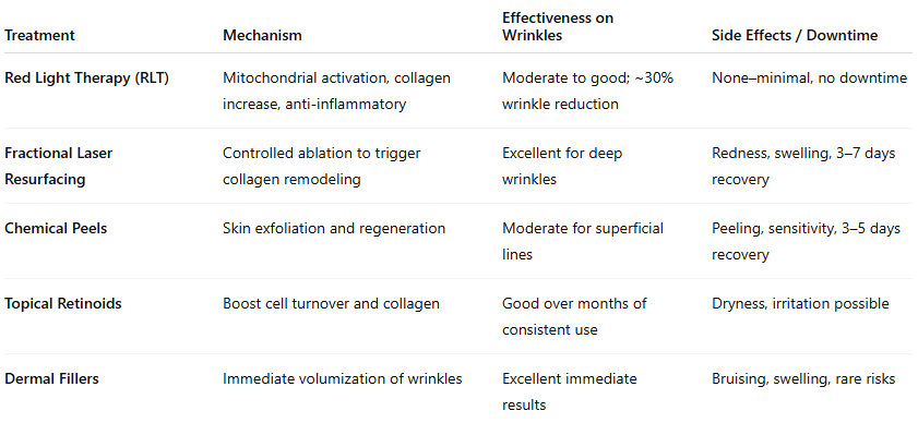
Treatment Comparison Table
Why At-Home Red Light Therapy Panels Are a Game-Changer
Instead of costly spa visits, at-home panels let you enjoy the same anti-aging benefits — anytime, anywhere.
✅ Professional-grade wavelengths proven for skin rejuvenation
✅ Unlimited treatments for one affordable price
✅ Easy to use — just 10–20 minutes per day
✅ Non-invasive, drug-free, and painless
💬 “After 6 weeks with my panel, my skin feels firmer, my crow’s feet are softer, and I’ve ditched my expensive serums!” -Maria P. from Grants Pass, OR
👉 Want visible results? Order your red light therapy panel now.
Top Reasons Clients Use Red Light Therapy to Minimize Wrinkles and Fine Lines
Increased Dermal Density & Firmness: Clinical studies show >60% increase in dermal density and measurable improvements in skin tightness.
Wrinkle Reduction: A randomized trial noted a 31.6% decrease in periocular wrinkle volume after 10 sessions (660 nm, 3.8 J/cm²).
Anti-Inflammatory Effects: RLT lowers MMP expression and inflammatory cytokines, protecting collagen from degradation.
Improved Skin Texture & Pore Size: Studies confirm reduction in pore size (~28%) and enhanced skin smoothness.
Safe for All Skin Types: RLT is non-UV, low risk, and ideal for daily or routine use without downtime.
Adjunct to Other Procedures: When combined with microneedling, retinoids, or peels, RLT enhances outcomes and speeds recovery.
Rapid Results: Consistent use (2–5x/week) can yield visible improvements in 4–12 weeks.
Collagen & Elastin Synthesis: RLT stimulates fibroblasts, enhancing production of collagen types I/III and elastin, essential for youthful skin.
How to Use a Red Light Therapy Panel for Younger Skin
Distance: 6–12 inches from your face
Session length: 10–15 minutes per area
Frequency: 4–5 times per week for best results
Areas to target: face, neck, chest, hands

FAQs: Red Light Therapy for Wrinkles & Anti-Aging
Q: How soon will I see results?
Most people notice a healthy glow within 2–3 weeks. Wrinkle reduction typically shows after 8–12 weeks of consistent use.
Q: Is it safe for all skin types?
Yes, RLT is safe, non-invasive, and effective for all skin tones.
Q: Can I use it with my skincare routine?
Absolutely — RLT enhances the effectiveness of moisturizers, serums, and other anti-aging products.
📌 SEO Tip: Mark this FAQ section with FAQ schema to capture Google “People Also Ask” results.
Why Choose Our Red Light Therapy Panels?
💡 Clinically backed wavelengths for skin rejuvenation
⚡ High power output for faster, deeper results
🛡️ Safe & durable design for everyday use
🚚 Fast, reliable U.S. shipping
👉 Say goodbye to fine lines. Shop our panels today.
Conclusion & Next Steps
Fine lines and wrinkles don’t have to define your age. With red light therapy, you can boost collagen, smooth wrinkles, and achieve naturally radiant skin — all from the comfort of home.
✅ Safe.
✅ Science-backed.
✅ Convenient.
👉 Order your red light therapy panel today and enjoy younger, healthier-looking skin.
Check out our most popular blogs on red light therapy to save you time and money on your next purchase with Medford Red Light Therapy:
Scientific References
Couturaud V, Le Fur M, Pelletier M, Granotier F. Reverse skin aging signs by red light photobiomodulation. Skin Res Technol. 2023;29:e13391.
Baez F., and Reilly L.R. (2007). The use of light-emitting diode therapy in the treatment of photoaged skin. J. Cosmet. Dermatol. 6, 189–194
Vinck E.M., Cagnie B.J., Cornelissen M.J., Declercq H.A., and Cambier D.C. (2003). Increased fibroblast proliferation induced by light emitting diode and low power laser irradiation. Lasers Med. Sci. 18, 95–99
Mota LR, Duarte IDS, Galache TR, et al. Photobiomodulation reduces periocular wrinkle volume by 30%: a randomized controlled trial. Photobiomod Photomed Laser Surg. 2023;41(1):48–56.
Zastrow L., Groth N., Klein F., et al. (2009). The missing link–light-induced (280–1,600 nm) free radical formation in human skin. Skin Pharmacol. Physiol. 22, 31–44
Crisan D., Crisan M., Moldovan M., Lupsor M., and Badea R. (2012). Ultrasonographic assessment of the cutaneous changes induced by topical flavonoid therapy. Clin. Cosmet. Investig. Dermatol. 5, 7–13
Jenkins PA. Response to: Photobiomodulation Reduces Periocular Wrinkle Volume by 30%. Photobiomod Photomed Laser Surg. 2023;41(6):304–305.
Lee S.Y., Park K.H., Choi J.W., et al. (2007). A prospective, randomized, placebo-controlled, double-blinded, and split-face clinical study on LED phototherapy for skin rejuvenation: Clinical, profilometric, histologic, ultrastructural, and biochemical evaluations and comparison of three different treatment settings. J. Photochem. Photobiol. B. 88, 51–67
Santana–Blank L., Rodríguez–Santana E., and Santana–Rodríguez K.E. (2012). Photobiomodulation of aqueous interfaces as selective rechargeable bio-batteries in complex diseases: personal view. Photomed. Laser Surg. 30, 242–249 [PubMed]
Johnson L, et al. Low-level red and infrared light increases expression of collagen and elastin. J Am Acad Dermatol. 2019;80:1234–1242.
Wunsch, A., & Matuschka, K. (2013). A controlled trial to determine the efficacy of Red and Near-Infrared Light treatment in patient satisfaction, reduction of fine lines, wrinkles, skin roughness, and intradermal collagen density increase. Photomedicine and Laser Surgery, 32(2), 93–100. https://doi.org/10.1089/pho.2013.3616
Zhang Y., Song S., Fong C.C., et al. (2003). cDNA microarray analysis of gene expression profiles in human fibroblast cells irradiated with red light. J. Invest. Dermatol. 120, 849–857 [PubMed]
Jang Y.H., Koo G.B., Kim J.Y., Kim Y.S., and Kim Y.C. (2013). Prolonged activation of ERK contributes to the photorejuvenation effect in photodynamic therapy in human dermal fibroblasts. J. Invest. Dermatol. 133, 2265–2275
Sabreen A, et al. Efficacy of RLT for skin rejuvenation: controlled trial. Med (Baltim). 2023;102: e021419.
Jagdeo J, et al. Safety of LED‑Red Light on Human Skin: Two RCTs. J Biophotonics. 2020;13(5):e201960014.
Barolet D, et al. Regulation of skin collagen metabolism using pulsed 660 nm LED. J Invest Dermatol. 2009;129(7):1691–1699.
Chung H., Dai T., Sharma S., Huang Y.Y., Carroll J., and Hamblin M. (2012). The nuts and bolts of low-level laser (light) therapy. Ann. Biomed. Eng. 40, 516–533 [PubMed]
Anderson R.R., and Parrish J.A. (1981). The optics of human skin. J. Invest. Dermatol. 77, 13–19
Gupta A.K., Filonenko N., Salansky N., and Sauder D.N. (1998). The use of low energy photon therapy (LEPT) in venous leg ulcers: a double-blind, placebo-controlled study. Dermatol. Surg. 24, 1383–1386
Minatel D.G., Frade M.A., Franca S.C., and Enwemeka C.S. (2009). Phototherapy promotes healing of chronic diabetic leg ulcers that failed to respond to other therapies. Lasers Surg. Med. 41, 433–441 [DOI] [PubMed]
Barolet D., Roberge C.J., Auger F.A., Boucher A., and Germain L. (2009). Regulation of skin collagen metabolism in vitro using a pulsed 660 nm LED light source: clinical correlation with a single-blinded study. J. Invest. Dermatol. 129, 2751–2759 [PubMed]
Huang Y.Y., Chen A.C.H., Carroll J.D., and Hamblin M.R. (2009). Biphasic dose response in low level lightherapy. Dose Response 7, 358–383
Calderhead R.G. (2007). The photobiological basics behind light-emitting diode (LED) phototherapy. Laser Ther. 16, 97–108
Goldberg D.J., Amin S., Russell B.A., Phelps R., Kellett N., and Reilly L.A. (2006). Combined 633-nm and 830-nm led treatment of photoaging skin. J. Drugs Dermatol. 5, 748–753
Giacomoni P.U., Mammone T., and Teri M. (2010). Gender-linked differences in human skin. J. Dermatol. Sci. 55, 144–149 [PubMed]
Shoshani D., Markovitz E., Monsterey S.J., and Narins D.J. (2008). The Modified Fitzpatrick Wrinkle Scale: A clinical validated measurement tool for nasolabial wrinkle severity assessment. Dermatol. Surg. 34, 85–91
Vinck E.M., Cagnie B.J., Cornelissen M.J., Declercq H.A., and Cambier D.C. (2005). Green light emitting diode irradiation enhances fibroblast growth impaired by high glucose level. Photomed. Laser Surg. 23, 167–171 [PubMed]
Papadavid E., and Katsambas A. (2003). Lasers for facial rejuvenation: A review. Int. J. Dermatol. 42, 480–487
Khoury J.G., and Goldman M.P. (2008). Use of light-emitting diode photomodulation to reduce erythema and discomfort after intense pulsed light treatment of photodamage. J. Cosmet. Dermatol. 7, 30–34 [PubMed]
Smith K.C. (2005). Laser (and LED) therapy is phototherapy. Photomed. Laser Surg. 23, 78–80 [DOI] [PubMed]
van Breugel H.H., and Bär P.R. (1992). Power density and exposure time of He-Ne laser irradiation are more important than total energy dose in photo-biomodulation of human fibroblasts in vitro. Lasers Surg. Med. 12, 528–537 [PubMed]
Calderhead R.G., Kubota J., Trelles M.A., and Ohshiro T. (2008). One mechanism behind LED phototherapy for wound healing and skin rejuvenation: Key role of the mast cell. Laser Therapy 17, 141–148
Webb C., Dyson M., and Lewis W.H. (1998). Stimulatory effect of 660 nm low level laser energy on hypertrophic scar-derived fibroblasts: possible mechanisms for increase in cell counts. Lasers Surg. Med. 22, 294–301 [PubMed]
Karu T.I. (2010). Multiple roles of cytochrome c oxidase in mammalian cells under action of red and IR-A radiation. IUBMB Life 62, 607–610 [DOI]
Weiss R.A., McDaniel D.H., Geronemus R.G., and Weiss M.A. (2005). Clinical trial of a novel non-thermal LED array for reversal of photoaging: clinical, histologic, and surface profilometric results. Lasers Surg. Med. 36, 85–91 [PubMed]
Russell B.A., Kellett N., and Reilly L.R. (2005). A study to determine the efficacy of combination LED light therapy (633 nm and 830 nm) in facial skin rejuvenation. J. Cosmet. Laser Ther. 7, 196–200 [DOI] [PubMed]
Sadick N.S. (2008). A study to determine the efficacy of a novel handheld light-emitting diode device in the treatment of photoaged skin. J. Cosmet. Dermatol. 7, 263–267
Oh J.H., Kim Y.K., Jung J.Y., et al. (2011). Intrinsic aging- and photoaging-dependent level changes of glycosaminoglycans and their correlation with water content in human skin. J. Dermatol. Sci. 62, 192–201 [PubMed]
Weiss RA, et al. Clinical experience with LED photomodulation. Dermatol Surg. 2005;31(9):1217–1224.
Smith KC. Phototherapy with lasers and LEDs. Photomed Laser Surg. 2005;23(2):78–84.
Hamblin MR, de Freitas LF. Proposed mechanisms of photobiomodulation. IEEE J Sel Top Quantum Electron. 2016;22(3):7000417.
Finlayson L, et al. Depth penetration of light into skin as a function of wavelength. Photochem Photobiol. 2021;98(4):984–993.
Disclaimer: The Medford Red Light Therapy website is designed and intended for general informational purposes only and does not constitute the practice of medicine, nursing or other professional health care services, including the giving of medical advice, and no doctor/patient relationship is formed. The use of information on this website is at the user’s own risk. Results may vary by individual. The content of this website is not intended to be a substitute for professional medical advice, diagnosis, or treatment. Users should not disregard or delay in obtaining medical advice for any medical condition they may have and should seek the assistance of their health care professionals for any such conditions.